By CARE Fertility and Women's Health
July 29, 2025
Introduction
Infertility, a condition affecting a significant portion of the global population, is often perceived as an intractable diagnosis, leaving many couples feeling powerless. However, a growing body of scientific research demonstrates that the underlying quality of the gametes—the egg (oocyte) and the sperm (spermatozoon)—is a key determinant of reproductive success and is, to a significant degree, modifiable. The journey to conception is fundamentally a story of cellular health. The viability of an embryo is contingent on the biological competence of the gametes that form it. Factors that were once considered immutable, such as age-related decline, are now understood through a cellular and molecular lens, revealing pathways for intervention.
This report will move beyond simplistic advice to provide a definitive, research-backed guide to understanding and enhancing gamete quality. We will deconstruct the intricate science of oocyte and spermatozoon biology, critically evaluate the evidence for nutritional, lifestyle, and environmental interventions, and synthesize these findings into a unified strategy for individuals and couples seeking to proactively improve their reproductive potential. This document is designed not as a replacement for medical consultation, but as a comprehensive resource to empower readers for more informed, productive discussions with their reproductive specialists, transforming them from passive recipients of care into active partners in their fertility journey.
Part I: The Oocyte - The Cellular Engine of New Life
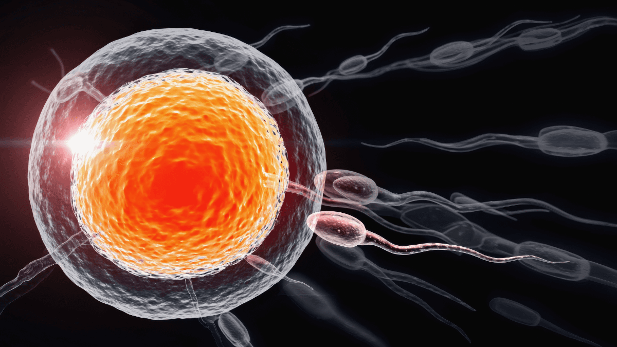
The female gamete, the oocyte, is a remarkable cell. It is the largest cell in the human body and carries not only the maternal genetic blueprint but also the complete cellular machinery required to initiate and sustain the first several days of embryonic life. The quality of this single cell is arguably the most critical factor in determining the outcome of a conception attempt. Understanding the biological principles that define oocyte quality is the first step toward influencing it. The central theme that emerges from decades of research is that oocyte competence is fundamentally a story of cellular energy and the integrity of its immediate environment.
Section 1: The Biology of Oocyte Quality
The Powerhouse of the Egg: The Critical Role of Mitochondria
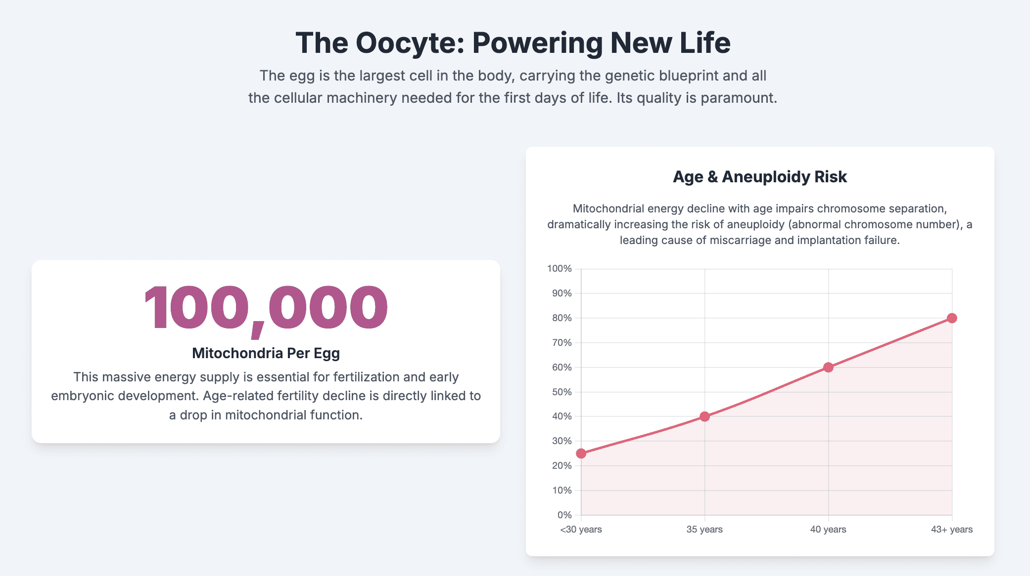
The oocyte's capacity to support life is directly tied to its energy supply. To this end, it is equipped with an unparalleled number of mitochondria—the powerhouses of the cell. A fully grown human oocyte contains approximately 100,000 mitochondria, the largest complement of any cell in the body, a biological testament to the immense energy required for the monumental tasks of maturation, fertilization, and early embryonic development.¹ These maternally inherited mitochondria are the sole source of adenosine triphosphate (ATP), the cell's energy currency, for the embryo until it reaches the blastocyst stage, as mitochondrial replication is suppressed following fertilization.²
Oocyte mitochondria are unique. In their immature state, they are spherical, possess few internal folds (cristae), and are bioenergetically "silent," producing ATP at a slow rate.² This quiescent state is theorized to be an evolutionary strategy to protect the mitochondrial genome (mtDNA) from oxidative damage. Because these mitochondria will be passed down to the embryo, minimizing the accumulation of mutations is paramount.² As the oocyte matures, these mitochondria undergo a crucial redistribution. They transition from a homogenous, scattered pattern throughout the cytoplasm to a clustered aggregation around the meiotic spindle, the apparatus that separates chromosomes.⁴ This precise movement, orchestrated by the cell's cytoskeleton, is essential for providing a localized, high-energy environment for the critical events of meiosis. A failure of this mitochondrial redistribution is a well-established marker of poor oocyte quality and can lead to developmental arrest due to an inability to supply ATP where it is most needed.⁴
Oocyte Aging and Aneuploidy: The Energy Deficit Hypothesis
The well-documented decline in female fertility with advancing age is intrinsically linked to a decline in mitochondrial function.¹ As a woman ages, her oocytes—which have been arrested in development since before her birth—suffer from accumulating cellular damage.⁵ The mitochondria within these aging oocytes become less efficient, leading to a significant decrease in their ability to synthesize ATP.¹
This resulting energy deficit has devastating consequences for the oocyte's genetic integrity. The segregation of chromosomes during meiosis is a highly energy-dependent process. Insufficient ATP compromises the formation and function of the meiotic spindle, leading to errors in chromosome separation known as meiotic nondisjunction. This is the primary mechanism responsible for the dramatic increase in aneuploidy (an abnormal number of chromosomes) observed in oocytes from women of advanced maternal age.¹ An embryo with an incorrect number of chromosomes is the leading cause of implantation failure and early pregnancy loss.
This functional decline is compounded by physical damage to the mitochondrial genome itself. Mitochondrial DNA (mtDNA) is a small, circular molecule containing 37 genes essential for the respiratory chain.¹ Unlike nuclear DNA, mtDNA lacks protective histone proteins and is located in close proximity to the electron transport chain, the primary site of ATP production and also the main source of damaging reactive oxygen species (ROS). Over decades, this constant exposure leads to the accumulation of mtDNA mutations and deletions, such as the well-studied 4977-bp "common deletion".¹ This damage creates a vicious cycle: damaged mtDNA impairs mitochondrial function, which reduces energy production and can increase ROS leakage, leading to further mtDNA damage.¹ Clinical studies have solidified this connection, demonstrating that altered levels of mtDNA in embryos are directly associated with female age, aneuploidy, and provide an independent predictor of an embryo's potential to implant.⁶ This establishes a clear, evidence-based cascade: advancing age leads to cumulative oxidative stress, which causes mitochondrial dysfunction and mtDNA damage. This, in turn, results in decreased ATP production, impaired meiotic spindle function, chromosomal errors, and ultimately, infertility or miscarriage.
Clinical Assessment of Oocyte Quality: From Morphology to AI
In the clinical setting of in vitro fertilization (IVF), embryologists must select the most promising oocytes for fertilization. The traditional and most widely used method for this is morphological assessment, guided by consensus criteria developed by leading reproductive medicine societies like the European Society of Human Reproduction and Embryology (ESHRE) and Alpha Scientists in Reproductive Medicine.⁷ According to these guidelines, a "good quality" oocyte is typically defined by a collection of visual features: a clear cytoplasm with fine, homogenous granularity; a normal round shape; a small, clear perivitelline space (the area between the oocyte and its outer shell); and an intact, non-fragmented first polar body (a small cellular byproduct of meiosis).⁸
However, a critical point for patients to understand is that the clinical utility of this visual checklist is surprisingly limited. The physical appearance of an oocyte is often a poor proxy for its true developmental competence. A 2023 presentation at the ESHRE annual meeting revealed that manual oocyte quality scoring performed by trained embryologists does not reliably correlate with whether that oocyte will successfully develop into a blastocyst-stage embryo.⁹ The scoring is inherently subjective, and most of the noted abnormalities (dysmorphisms) are rare and do not consistently predict poor outcomes.
The future of oocyte assessment is shifting toward more objective, data-driven methods. The same ESHRE presentation highlighted an artificial intelligence (AI) image analysis tool, MAGENTA, which was trained to analyze oocyte images for parameters that may be imperceptible to the human eye. The AI-generated scores, unlike the human scores, did significantly correlate with an oocyte's potential to form a blastocyst.⁹ This represents a paradigm shift, suggesting that the most critical markers of quality are likely subcellular—related to factors like mitochondrial distribution and metabolic health—rather than overt morphological flaws. This knowledge can be empowering for patients who may feel distressed by an embryology report noting "poor morphology," as it underscores that the visual report is not the final word on an oocyte's potential.
The primacy of intrinsic biological quality over external factors is further highlighted by oocyte cryopreservation (egg freezing) outcomes. While vitrification technology has improved dramatically, success rates remain profoundly tied to the age at which the oocytes were frozen.¹⁰ ASRM guidelines note that live birth rates are improved when oocytes are cryopreserved at a younger age. The technology can preserve an oocyte in its current state but cannot reverse the intrinsic, age-related decline in its cellular machinery.¹⁰
Section 2: Medical Conditions Impacting the Ovarian Environment
The oocyte does not develop in a vacuum. It is nurtured within a follicle, bathed in follicular fluid, and supported by a community of surrounding cumulus and granulosa cells.¹ The health of this "ovarian microenvironment" is as important as the intrinsic quality of the oocyte itself. Systemic medical conditions can profoundly alter this environment, creating suboptimal conditions for oocyte maturation.
Polycystic Ovary Syndrome (PCOS): A Paradox of Quantity Over Quality
Polycystic Ovary Syndrome (PCOS) is one of the most common endocrine disorders in women of reproductive age and a leading cause of anovulatory infertility.¹¹ It is diagnosed based on a combination of criteria, including irregular or absent ovulation, clinical or biochemical signs of high androgen levels (hyperandrogenism), and the presence of polycystic ovarian morphology on ultrasound.¹³
During IVF, women with PCOS often exhibit a robust response to ovarian stimulation, producing a large number of oocytes. This apparent abundance, however, creates a paradox of quantity versus quality.¹⁴ The core issue in PCOS is the adverse follicular microenvironment created by the syndrome's underlying metabolic and endocrine disruptions. The hormonal milieu is characterized by elevated levels of luteinizing hormone (LH), hyperandrogenism, and often, insulin resistance.¹⁶ These systemic imbalances translate directly to the follicular fluid. For instance, studies have found that the follicular fluid of women with PCOS contains increased levels of free fatty acids, which are positively correlated with poor subsequent embryo morphology and fragmentation.¹⁶
Systematic reviews and meta-analyses paint a complex but consistent picture. PCOS patients undergoing IVF may experience higher cycle cancellation rates and have lower fertilization rates per oocyte compared to women without PCOS.¹¹ Yet, they often achieve comparable overall clinical pregnancy and live birth rates. This is likely because the sheer number of oocytes retrieved compensates for the lower efficiency of each individual oocyte.¹¹ This highlights that the fundamental challenge in PCOS-related infertility is not a lack of eggs, but a lower competence of each egg due to its developmental environment. Furthermore, evidence suggests that different PCOS phenotypes—combinations of the diagnostic criteria—may have varying impacts. The combination of hyperandrogenism and chronic anovulation appears to be particularly detrimental to oocyte competence.¹⁴
Endometriosis: The Impact of Systemic and Local Inflammation
Endometriosis, a condition where endometrial-like tissue grows outside the uterus, impairs fertility through multiple pathways. While it can cause anatomical distortions that block the fallopian tubes, a significant and often overlooked mechanism is its negative impact on oocyte quality.¹⁷ The disease establishes a chronic pro-inflammatory state within the pelvic cavity and, when endometriomas (cysts) are present, within the ovary itself.¹⁹
This pervasive inflammation leads to a hostile follicular microenvironment. The follicular fluid becomes saturated with inflammatory mediators and reactive oxygen species, creating a state of high oxidative stress.¹⁷ This toxic milieu can directly damage the developing oocyte, leading to altered morphology, a lower cytoplasmic mitochondrial content, and an increased risk of meiotic abnormalities and chromosomal instability.¹⁷ Some research suggests the primary damage may be to the surrounding granulosa cells, which then fail in their crucial role of supporting the oocyte's development.¹⁹
The scientific literature on this topic presents a fascinating conflict, which itself reveals a deeper truth. Some systematic reviews conclude that oocytes from women with endometriosis are less likely to mature properly in vitro and result in lower quality embryos.¹⁷ This view is supported by oocyte donation studies: when healthy donor eggs are transferred to women with endometriosis, pregnancy rates are often normal, strongly suggesting that the primary fertility defect lies within the patient's own oocytes.¹⁷ Conversely, a large 2021 meta-analysis concluded that from a purely morphological perspective, endometriosis does not compromise embryo quality.²³ This apparent contradiction is not a simple disagreement; it suggests that the damage inflicted by endometriosis may be at a molecular or metabolic level—such as impaired mitochondrial function—that is not visible during standard microscopic grading. This reinforces the limitations of visual assessment and points to a more subtle, but no less significant, form of damage. The inconsistency in findings may also be due to variations in disease severity, location of lesions, and the frequent co-existence of adenomyosis, which is often not separated out in studies.²¹
Section 3: Evidence-Based Nutraceuticals for Enhancing Female Fertility
Given that oocyte quality is tied to cellular energy and the ovarian environment, it is logical that interventions targeting these pathways could be beneficial. A number of nutraceuticals have been studied for this purpose, with the strongest evidence emerging for those that address specific, well-understood biological mechanisms. Effective supplementation is not a one-size-fits-all approach but is instead mechanism-based and targeted to the individual's underlying pathophysiology.
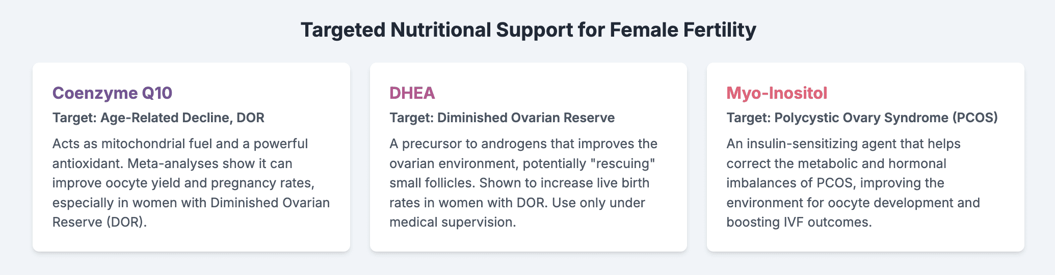
Coenzyme Q10 (CoQ10): Fueling the Cellular Engine
Mechanism: Coenzyme Q10 (CoQ10) is a vitamin-like substance that is a vital component of the mitochondrial electron transport chain, the cellular machinery responsible for generating ATP. It also functions as a powerful, lipid-soluble antioxidant, protecting mitochondrial membranes and mtDNA from damage by reactive oxygen species.²⁴ This dual role in both energy production and antioxidant defense makes it an ideal candidate for directly addressing the core mechanism of oocyte aging: mitochondrial dysfunction.
Evidence: The rationale for CoQ10 supplementation is strongly supported by research. Studies in aged animal models have shown that CoQ10 can delay the depletion of the ovarian reserve and restore mitochondrial gene expression and activity in oocytes to levels seen in younger animals.²⁷ Human studies confirm that CoQ10 is naturally present in follicular fluid, and that higher levels are positively correlated with better oocyte and embryo quality.²⁵ Building on this, multiple meta-analyses of randomized controlled trials (RCTs) have linked CoQ10 supplementation with significant improvements in IVF outcomes, particularly for women with diminished ovarian reserve (DOR) or a history of poor ovarian response. These benefits include an increased number of retrieved oocytes, a higher number of top-quality embryos, and, most importantly, higher clinical pregnancy rates.²⁶
Dosage and Duration: A critical finding from the research is that the duration of supplementation is paramount. The development of an ovarian follicle from the early preantral stage to a mature, ovulatory follicle takes several months. A 2024 meta-analysis found a significant benefit in clinical pregnancy rates only when CoQ10 supplementation was continued for at least three months prior to the IVF cycle.²⁸ This aligns perfectly with the known timeline of folliculogenesis, indicating that short-term supplementation is unlikely to be effective. Dosages used in successful clinical trials typically range from 200 mg to 600 mg per day.²⁶
Dehydroepiandrosterone (DHEA): Improving the Ovarian Environment
Mechanism: Dehydroepiandrosterone (DHEA) is a mild adrenal steroid hormone that serves as a precursor to androgens like testosterone. Within the ovary, androgens are believed to play a crucial role in the early stages of follicular development, promoting the growth of small preantral and antral follicles and reducing the rate of follicular atresia (programmed cell death).³⁰ The leading hypothesis is that DHEA does not "rejuvenate" the oocytes themselves, but rather improves the "aging ovarian environment." By creating a more androgen-replete milieu, it may rescue small follicles that would otherwise perish, allowing them to grow and become available for recruitment during an IVF cycle.²⁷
Evidence: DHEA has been studied almost exclusively in women with DOR, who are characterized by a poor ovarian response to stimulation. The evidence, while still evolving, is promising. A 2021 systematic review and meta-analysis of nine RCTs concluded that DHEA supplementation in women with DOR or poor ovarian response led to a statistically significant increase in live birth rates and the number of retrieved oocytes.³¹ Other studies have similarly found that DHEA pretreatment can increase both oocyte and embryo yields, particularly in older patients and those classified under the POSEIDON group 4 criteria (older patients with low ovarian reserve markers).³² Some research even suggests DHEA may improve embryo quality by reducing aneuploidy rates.³⁰
Dosage and Considerations: The most common dosage regimen in clinical research is 75 mg per day, typically administered as 25 mg three times daily.²⁷ As with CoQ10, timing is important; supplementation for at least 6–8 weeks, and ideally for 3–4 months, prior to an IVF cycle is recommended to influence the cohort of developing follicles.²⁷ It is imperative that DHEA is used only under medical supervision. As an androgen, it can have side effects such as oily skin, acne, and hair growth, and its use should be monitored by a physician.
Myo-Inositol: Targeting Metabolic Health in PCOS
Mechanism: Myo-inositol is a member of the B-vitamin complex family and a key intracellular signaling molecule. It functions as a "second messenger" for critical hormones, including follicle-stimulating hormone (FSH) and, most importantly, insulin.³³ Many women with PCOS suffer from insulin resistance, a condition where cells do not respond efficiently to insulin, leading the pancreas to produce excess amounts of it (hyperinsulinemia). This hyperinsulinemia is a primary driver of the hyperandrogenism seen in PCOS. Myo-inositol acts as an insulin-sensitizing agent, improving the cell's glucose uptake and helping to normalize insulin levels.³⁴ By correcting the underlying metabolic dysfunction, it helps create a healthier and more balanced systemic and ovarian environment for follicular development.
Evidence: Given its targeted mechanism, myo-inositol is a cornerstone of nutraceutical management for PCOS. A 2024 joint position statement from several international expert groups recommended that myo-inositol be considered as a pretreatment strategy prior to ovarian stimulation in IVF, particularly for women with PCOS.³³ Clinical studies and meta-analyses have shown that it can improve the metabolic profile of women with PCOS, reduce insulin levels and hyperandrogenism, potentially reduce the required dose of gonadotropins during IVF, improve oocyte and embryo quality, and increase clinical pregnancy rates.³³
Dosage and Formulation: The ovary naturally maintains a specific ratio of myo-inositol to another isomer, D-chiro-inositol, of approximately 40:1. In PCOS, this ratio is often disrupted. Therefore, supplementation that provides these inositols in the physiological 40:1 ratio is often recommended to restore this balance.³⁴ A typical clinical dosage is 2 grams of myo-inositol (often with 50 mg of D-chiro-inositol) taken twice daily. The supplement is generally recognized as safe, with only minor gastrointestinal side effects at very high doses.³³
Part II: The Spermatozoon - A Journey of Maturation and Motility
While the oocyte provides the initial cellular infrastructure, the spermatozoon delivers the paternal genetic contribution and the spark that activates embryonic development. For decades, the focus of fertility evaluation was disproportionately on the female partner. However, it is now unequivocally established that male factors contribute to approximately half of all infertility cases.³⁶ The quality of sperm is the end-product of a long, complex, and vulnerable developmental process, and its proper assessment requires looking far beyond a simple count.
Section 4: The Biology of Sperm Quality
The Spermatogenesis Cycle: A ~74-Day Production Timeline
Unlike the female, who is born with a finite supply of oocytes, the male is a continuous production factory for sperm. This process, known as spermatogenesis, is a remarkable biological assembly line that transforms a simple stem cell into a highly specialized, motile cell capable of fertilization. The entire developmental journey, from a spermatogonial stem cell to a mature spermatozoon ready for ejaculation, takes approximately 74 days, with some estimates extending to 120 days when including transport through the ductal system.³⁷
This timeline is of profound clinical and practical importance. It establishes a clear, biologically-mandated window for intervention. Any lifestyle change, nutritional supplement, or medical treatment aimed at improving sperm quality will not be reflected in the ejaculate for nearly three months. This knowledge is crucial for managing expectations and encouraging the sustained effort required to see measurable results. A weekend of healthy living will not change a semen analysis; a season of it might.
Deconstructing the Semen Analysis: A Guide to the WHO 2021 Criteria
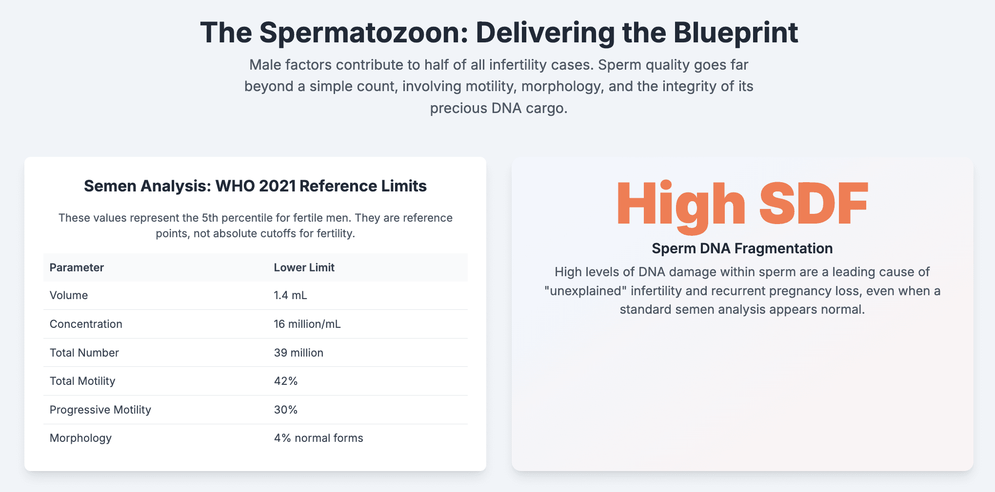
The cornerstone of the modern male fertility evaluation is the semen analysis (SA).⁴³ To ensure consistency and reliability across laboratories worldwide, the World Health Organization (WHO) publishes a detailed manual for the examination of human semen. The most current version is the 6th Edition, released in 2021, which provides updated, evidence-based procedures and new reference values derived from studies of fertile men.⁴⁴
A comprehensive semen analysis, according to ASRM and WHO guidelines, evaluates several key parameters⁴⁸:
Semen Volume: The total amount of fluid in the ejaculate.
Sperm Concentration (or Density): The number of sperm per milliliter of semen.
Total Sperm Number: The total number of sperm in the entire ejaculate (concentration multiplied by volume).
Sperm Motility: The percentage of sperm that are moving. The 2021 WHO criteria reintroduced a more detailed breakdown:
Progressive Motility (PR): Sperm moving actively, either in a straight line or in large circles. This is the most functionally important type of movement.
Non-Progressive Motility (NP): Sperm that show movement (e.g., twitching tails) but do not move forward.
Total Motility: The sum of progressive and non-progressive sperm (PR + NP).
Sperm Morphology: The size and shape of the sperm. It assesses the percentage of sperm that have a "normal" structure (typically an oval head and a single, long tail), as defects can impair the sperm's ability to navigate the female reproductive tract and penetrate the egg.⁵¹
Sperm Vitality: The percentage of sperm that are alive, determined by a membrane-integrity test. This is particularly important when motility is very low to distinguish between dead sperm and live but immotile sperm.⁵⁰
Other Parameters: The analysis also assesses pH, time to liquefaction, and the presence of white blood cells, which could indicate an infection.⁴⁹
It is absolutely crucial to understand how to interpret these results. The WHO provides "lower reference limits" (LRLs), which represent the 5th percentile of values from a population of men who recently fathered a child.⁴⁷ These are not absolute cutoffs for fertility or infertility. A man can have parameters below these limits and still be fertile, and conversely, a man with all "normal" parameters can be infertile.⁴³ Due to the high biological variability in semen parameters from day to day, guidelines from the American Urological Association (AUA) and ASRM strongly recommend that at least two semen analyses, ideally obtained at least a month apart, are performed before making any clinical judgments, especially if the first result is abnormal.⁴³
Beyond conception, semen quality is increasingly recognized as a biomarker for overall male health. Studies have linked low sperm counts to a higher prevalence of metabolic issues like increased body fat and high blood pressure, and one large, long-term study found that men with poor semen quality had a shorter life expectancy, suggesting that the factors affecting fertility may also impact systemic health.⁵⁴
Parameter | WHO 2021 Lower Reference Limit (5th Percentile, 95% CI) |
Semen Volume (mL) | 1.4 (1.3–1.5) |
Sperm Concentration (106 per mL) | 16 (15–18) |
Total Sperm Number (106 per ejaculate) | 39 (35–40) |
Total Motility (PR + NP, %) | 42 (40–43) |
Progressive Motility (PR, %) | 30 (29–31) |
Vitality (live spermatozoa, %) | 54 (50–56) |
Sperm Morphology (normal forms, %) | 4 (3.9–4.0) |
Data derived from the WHO laboratory manual for the examination and processing of human semen, 6th edition.47 |
Beyond the Basics: The Clinical Significance of Sperm DNA Fragmentation
A standard semen analysis provides critical information about the quantity and quality of the sperm cells—the "vehicles." However, it reveals nothing about the integrity of their precious cargo: the paternal DNA. Sperm DNA Fragmentation (SDF) refers to breaks or damage in the DNA strands contained within the sperm head. While the oocyte has some capacity to repair this damage after fertilization, extensive fragmentation can overwhelm its repair mechanisms.
High SDF is now a well-recognized, independent cause of male infertility that is not captured by a standard SA. It is strongly associated with lower rates of natural conception, reduced success in IVF and ICSI cycles, impaired embryo development, and a significantly increased risk of recurrent pregnancy loss (RPL).⁴³ This provides a potential explanation for many cases of "unexplained" infertility or recurrent miscarriage where the male partner has a "normal" semen analysis.
Reflecting its growing clinical importance, the joint AUA/ASRM guidelines recommend considering SDF testing for couples experiencing RPL (two or more losses).⁴³ However, it is not yet recommended as part of the routine initial evaluation for all infertile couples. Several laboratory techniques, such as the TUNEL assay or Sperm Chromatin Dispersion (SCD) test, can be used to measure the percentage of sperm with fragmented DNA.⁴⁶
Section 5: Key Pathologies and Detriments to Sperm Health
While some cases of male infertility are idiopathic (of unknown cause), many stem from identifiable conditions or exposures. These factors often converge on a final common pathway of cellular damage, compromising the delicate process of spermatogenesis.
Varicocele: The Most Common Correctable Cause
A varicocele is an abnormal dilation of the veins within the pampiniform plexus of the scrotum, analogous to a varicose vein in the leg. It is the most common identifiable and, importantly, correctable cause of male infertility, found in up to 40% of men with primary infertility and up to 81% with secondary infertility.²²
The exact pathophysiology of how a varicocele impairs sperm production is multifactorial and still under investigation, but several key mechanisms are proposed. The leading theories include scrotal hyperthermia (the pooling of venous blood raises testicular temperature above the optimal level for spermatogenesis), the retrograde flow (reflux) of toxic metabolites from the adrenal gland and kidney down to the testis, and testicular hypoxia (low oxygen).⁵⁶ However, a central and unifying mechanism appears to be a dramatic increase in local oxidative stress.²²
Treatment via surgical repair (varicocelectomy) or embolization is recommended by AUA/ASRM for men who have a clinically palpable varicocele, documented infertility, and abnormal semen parameters, provided the female partner has normal or correctable fertility.⁵⁹ Numerous studies and systematic reviews have confirmed that varicocele repair is associated with significant improvements in key semen parameters, particularly sperm concentration and motility.⁵⁶ Furthermore, the degree of improvement often correlates directly with the preoperative grade (severity) of the varicocele, with higher-grade varicoceles showing a greater response to treatment.⁵⁷
Oxidative Stress: The Pervasive Threat to Sperm Integrity
Oxidative stress (OS) is a state of cellular imbalance where the production of damaging reactive oxygen species (ROS) overwhelms the body's protective antioxidant defenses. Spermatozoa are uniquely vulnerable to OS for two main reasons: their plasma membranes are rich in polyunsaturated fatty acids, which are easily oxidized, and they possess very little cytoplasm, which limits their intrinsic antioxidant capacity.⁶⁰
Excessive ROS, generated by sources like leukocytes (white blood cells) in the semen, infections, or environmental toxins, wreaks havoc on sperm. It attacks the sperm membrane, impairing its fluidity and function, which is critical for motility and fertilization. Most critically, ROS directly attacks the DNA within the sperm head, causing single- and double-strand breaks. This damage is the primary cause of high sperm DNA fragmentation (SDF).⁶⁰
The concept of oxidative stress is not just another factor in male infertility; it is the central biochemical pathway through which many different negative inputs exert their damage. Pathologies like varicocele²², systemic conditions like obesity⁶², psychological stress⁶³, and lifestyle choices like alcohol consumption⁶⁴ all contribute to increased seminal OS. This understanding provides a powerful and unifying rationale for the use of antioxidant therapies as a broad-spectrum strategy to protect sperm and improve male reproductive potential.
Section 6: Evidence-Based Nutraceuticals for Enhancing Male Fertility
Given the central roles of energy metabolism and oxidative stress in sperm health, it is logical that nutritional supplements targeting these pathways could offer therapeutic benefits. Research has identified several key compounds that have been shown in clinical trials and meta-analyses to improve semen parameters.
The Antioxidant Arsenal: L-Carnitine, CoQ10, and Vitamins
A primary strategy for combating oxidative stress-induced sperm damage is to bolster the body's antioxidant defenses through supplementation.
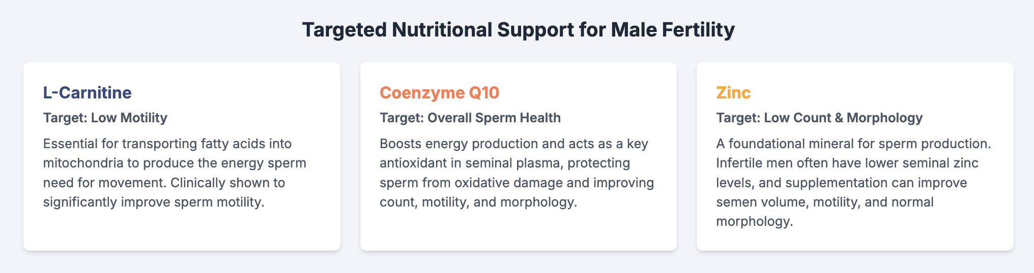
L-Carnitine: This amino acid derivative plays a pivotal role in transporting fatty acids into the mitochondria for energy production, a process that is absolutely essential for sperm motility. It is highly concentrated in the epididymis, where sperm undergo final maturation.⁶⁵ Multiple systematic reviews and meta-analyses have concluded that supplementation with L-carnitine (often in combination with L-acetyl-carnitine) can significantly improve key sperm parameters, most notably sperm motility and concentration, and may even increase pregnancy rates.³⁶
Coenzyme Q10: Much like in the oocyte, CoQ10 serves a dual function in sperm as a critical component of the mitochondrial respiratory chain for energy production and as a potent antioxidant. Studies have shown that CoQ10 supplementation can improve sperm count, motility, and morphology.⁶⁶ By boosting antioxidant capacity within the seminal plasma, it also helps protect sperm DNA from oxidative damage, which may lead to healthier embryo development.⁶⁷
General Antioxidants: A broader body of evidence supports the use of combined antioxidant formulas. Systematic reviews have consistently concluded that supplementation with a range of antioxidants—including Vitamin E, Vitamin C, and N-acetyl cysteine (NAC)—appears to have a favorable effect on semen parameters.⁶⁰ Some studies have even linked antioxidant therapy to improved outcomes in assisted reproductive technology (ART) and higher live-birth rates, although a clear consensus on the single most effective formula or dosage remains elusive.⁶⁰
Essential Minerals: The Nuanced Roles of Zinc and Selenium
Micronutrients are also critical for healthy sperm production, but the evidence highlights the importance of balance.
Zinc: This essential trace mineral is fundamental to the male reproductive system. Studies have shown that infertile men often have significantly lower concentrations of zinc in their seminal plasma compared to fertile controls. A 2016 meta-analysis of 20 studies found that zinc supplementation was associated with statistically significant increases in semen volume, sperm motility, and the percentage of sperm with normal morphology.⁴²
Selenium: The evidence for selenium supplementation is more complex and serves as an important cautionary tale. While selenium is a crucial component of antioxidant enzymes like glutathione peroxidase, which protects sperm from oxidative damage, the clinical trial data is conflicting. A recent, large meta-analysis examining the effects of co-supplementation with selenium and vitamin E delivered a paradoxical result: while the combination improved sperm vitality (the percentage of live sperm) and normal morphology, it also simultaneously and significantly reduced semen volume and total sperm count.⁶⁸ This demonstrates that the biochemistry of the testis is a delicate balance. Simply flooding the system with a single type of antioxidant can have unintended negative consequences on other aspects of sperm production. This argues against the practice of megadosing single nutrients and underscores the importance of a balanced, comprehensive approach, ideally guided by a physician.
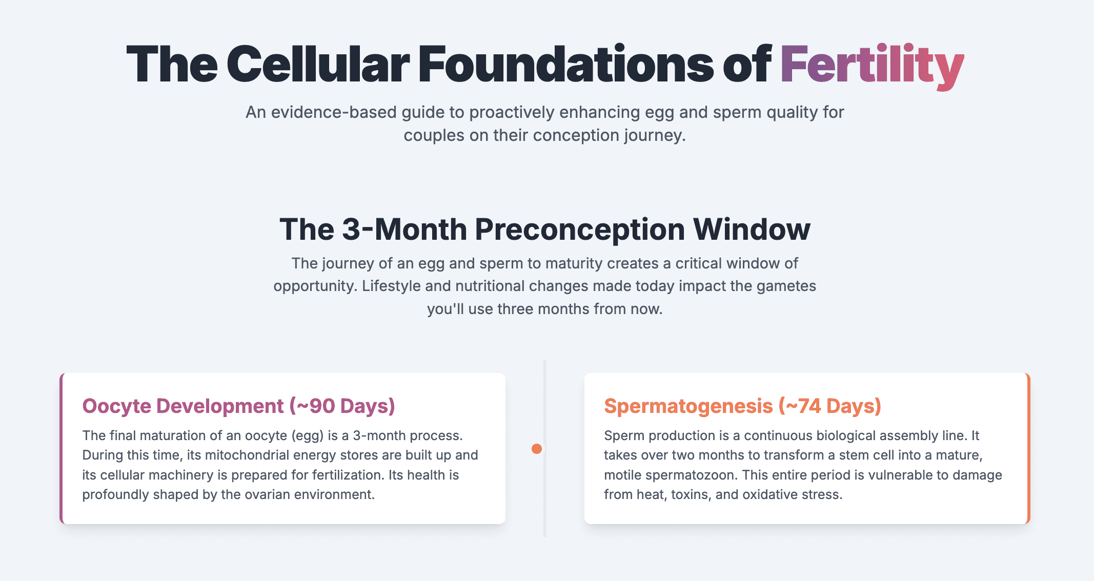
Part III: A Unified Approach - Shared Lifestyle and Environmental Factors
While infertility is often investigated by focusing on either the male or female partner, the reality is that a couple's health and habits are intertwined. Many of the most powerful modifiable factors that influence gamete quality are shared. Diet, weight, stress, and environmental exposures do not discriminate; they affect both partners, and addressing them as a team can be one of the most effective strategies for improving fertility. Fertility is a team sport, and shared habits have a profound impact on shared outcomes. The ~3-month period corresponding to both a full spermatogenesis cycle and a critical window of oocyte development represents a crucial window of opportunity for couples to implement these changes together.
Section 7: The Foundational Pillars: Diet and Weight
The Mediterranean Diet: An Anti-Inflammatory Blueprint for Couple Fertility
A growing body of evidence points to the profound impact of dietary patterns on reproductive health. The Mediterranean Diet (MD), in particular, has emerged as a powerful, evidence-based nutritional strategy for enhancing fertility. Characterized by a high intake of fruits, vegetables, legumes, nuts, whole grains, fish, and olive oil, and a low intake of red and processed meats, sweets, and ultra-processed foods, the MD is naturally rich in vitamins, minerals, fiber, and potent anti-inflammatory and antioxidant compounds.⁶⁹
The beneficial mechanism is clear: a common factor underlying many cases of infertility in both sexes is a state of chronic, low-grade inflammation and oxidative stress.⁶⁹ The MD directly counteracts this pathology.
For Men: Systematic reviews and meta-analyses have shown that higher adherence to a healthy dietary pattern like the MD is associated with significantly improved sperm quality, including higher sperm concentration, greater total sperm count, and better progressive motility.⁷¹
For Women: For the female partner, higher adherence to the MD has been linked in multiple studies to improved outcomes with assisted reproductive technology, including higher rates of clinical pregnancy and live birth.⁷³ The diet's rich array of antioxidants and vitamins may contribute directly to better oocyte quality, while its anti-inflammatory properties can create a more favorable endometrial environment for embryo implantation.⁷³ A 2023 systematic review further found that adherence to the MD during pregnancy may lower the risk of adverse outcomes like gestational diabetes and preterm birth.⁷⁴
The Impact of Body Mass Index (BMI): A Delicate Hormonal Balance
Body weight is a critical regulator of the endocrine system, and deviations from a healthy weight—in either direction—can severely disrupt the delicate hormonal balance required for fertility in both men and women.
For Women: Obesity (a high BMI) is a major risk factor for female infertility. It profoundly disrupts the hypothalamic-pituitary-ovarian (HPO) axis, the hormonal command chain that governs the menstrual cycle. This disruption frequently leads to anovulation.⁷⁵ Beyond ovulation, obesity creates a hostile metabolic environment that directly impairs oocyte quality. The mechanisms are multifaceted and include insulin resistance, which leads to compensatory hyperinsulinemia and subsequent ovarian hyperandrogenism, and altered levels of inflammatory adipokines (hormones secreted by fat cells, such as leptin) within the follicular fluid, creating a suboptimal milieu for oocyte maturation.⁷⁵ Consequently, obesity is linked to lower pregnancy rates and a higher risk of miscarriage.⁷⁵
For Men: Obesity is equally detrimental to male fertility. An increased BMI is consistently associated with poorer semen parameters, including lower sperm count, concentration, and motility.⁶² Furthermore, obesity is a significant risk factor for elevated sperm DNA fragmentation.⁶² The mechanisms in men also involve hormonal disruption, primarily lower testosterone and higher estrogen levels (due to the aromatization of testosterone to estrogen in excess adipose tissue), as well as direct heat effects from scrotal fat, and a state of systemic inflammation and oxidative stress that damages developing sperm.⁶²
The consistent, parallel damage caused by lifestyle and environmental factors in both partners underscores the importance of a collaborative approach to preconception health.
Factor | Impact on Oocyte Quality | Impact on Sperm Quality | Evidence-Based Recommendation |
Diet | High adherence to a Mediterranean-style diet is associated with improved ART outcomes and may improve oocyte quality.73 | High adherence to a healthy diet is associated with improved sperm concentration, count, and motility.71 | Adopt a Mediterranean-style diet rich in fruits, vegetables, whole grains, legumes, fish, and healthy fats, while limiting red/processed meat and sugar.69 |
BMI | Obesity (high BMI) disrupts hormones, impairs oocyte maturation, and increases miscarriage risk.75 | Obesity (high BMI) is associated with lower testosterone, reduced sperm count and motility, and increased DNA fragmentation.62 | Achieve and maintain a healthy BMI (18.5-24.9). Modest weight loss in overweight/obese individuals can significantly improve fertility outcomes.62 |
Exercise | Moderate exercise reduces the risk of anovulatory infertility. Excessive, high-intensity exercise can disrupt ovulation.78 | Moderate exercise improves sperm parameters. Intensive training can worsen them.61 | Engage in moderate, regular physical activity (e.g., 30-60 minutes, 3-5 times per week). Avoid overtraining and excessive high-intensity exercise.61 |
Sleep | Poor sleep (<7 hours/night) is associated with reduced oocyte and embryo production during IVF.81 | Both short (<6 hours) and long (>9 hours) sleep durations are associated with lower sperm count, survival, and motility.82 | Aim for 7-9 hours of consistent, high-quality sleep per night. Maintain a regular sleep-wake schedule.81 |
Stress | Chronic stress is associated with poorer IVF outcomes, including fewer retrieved oocytes.83 | Chronic stress impairs testicular function, reducing testosterone and sperm quality, and can cause sexual dysfunction.63 | Implement stress-reduction techniques such as mindfulness, meditation, yoga, or therapy. The infertility journey itself is a major stressor that requires active management.63 |
Alcohol | Can disrupt hormones controlling ovulation and may damage egg quality via oxidative stress. Heavy use reduces ART success.84 | Heavy consumption reduces semen volume and can negatively impact hormones and sperm development. Abstinence can reverse some damage.64 | Minimize or eliminate alcohol consumption while trying to conceive. The safest approach for both partners is abstinence.85 |
Smoking | Increases miscarriage risk and accelerates ovarian aging.87 | Increases inflammatory cells in semen and is associated with poorer sperm quality in some meta-analyses.88 | Complete cessation of smoking for both partners is strongly recommended. There is no safe level of smoking for fertility.87 |
BPA | Linked to PCOS, meiotic errors, and impaired oocyte maturation.89 | Meta-analyses show a negative correlation with sperm concentration and total count.91 | Avoid plastic containers with recycling codes 3 or 7. Opt for glass, stainless steel, or BPA-free plastics. Minimize handling of thermal paper receipts.93 |
Phthalates | Disrupts folliculogenesis (egg development) and steroidogenesis (hormone production).94 | Robust evidence links exposure to reduced semen quality (especially from DEHP and DBP) and lower testosterone.95 | Avoid products with "fragrance" listed as an ingredient. Choose phthalate-free personal care products and avoid plastic food containers and wrap, especially for heating food.97 |
Pesticides | Associated with menstrual cycle disturbances and longer time-to-pregnancy.98 | Insecticide exposure is consistently associated with lower sperm concentration in meta-analyses.100 | Choose organic produce when possible, especially for items on the "Dirty Dozen" list. Wash all fruits and vegetables thoroughly. Avoid using pesticides in the home and garden.99 |
Section 8: Daily Habits and Their Reproductive Consequences
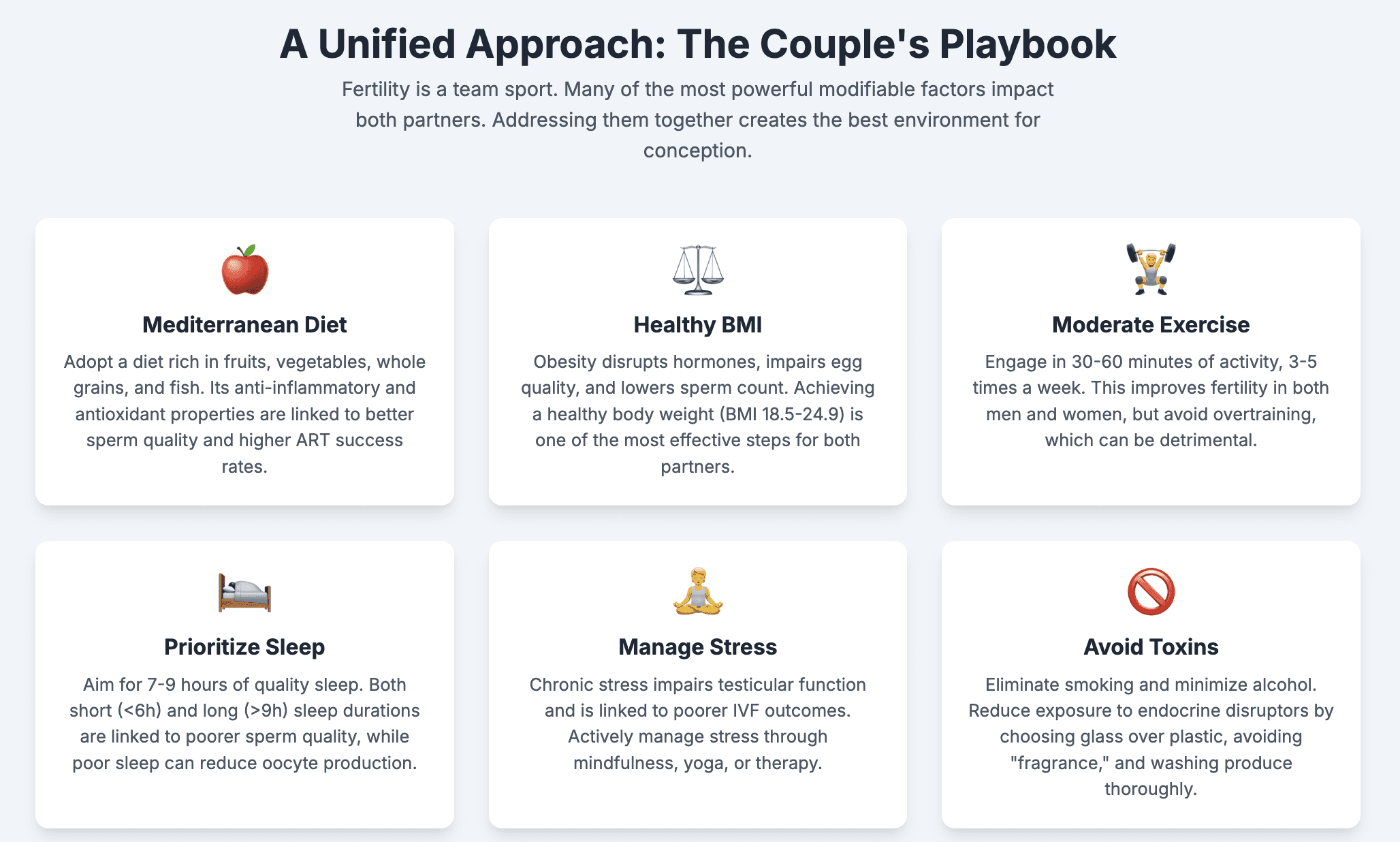
Exercise: Finding the Fertile Balance
Physical activity has a complex, U-shaped relationship with fertility: too little is detrimental, but too much can be as well. The key is finding a moderate, sustainable balance.
The Benefit of Moderation: Regular, moderate exercise is clearly beneficial for fertility. For women, a large cohort study found that vigorous activity of 30-60 minutes per day was associated with a reduced risk of ovulatory infertility.⁷⁸ For men, a network meta-analysis found that various forms of moderate exercise, including aerobic and resistance training, can significantly improve sperm volume, motility, and morphology.⁶¹
The Harm of Extremes: Conversely, excessive, high-intensity exercise is detrimental. In women, training for more than 60 minutes per day is associated with an increased risk of anovulation, likely due to the energetic stress disrupting the hypothalamic-pituitary-gonadal (HPG) axis.⁷⁸ In men, studies of athletes undergoing intensive training regimens show that it can worsen seminal parameters.⁸⁰ The goal is to be active, not to push the body to its physiological limits.
Sleep and Stress: The HPA-HPG Axis Connection
The body's reproductive axis (HPG) and its stress axis (hypothalamic-pituitary-adrenal, or HPA) are deeply and inversely connected. When the stress axis is chronically activated, the reproductive axis is often suppressed. This makes management of both stress and sleep fundamental to fertility.
Impact on Women: A systematic review of stress and IVF outcomes found a clear negative effect, with the most significant impact seen at the egg retrieval stage, where higher stress was associated with a lower number of oocytes collected.⁸³ Sleep is equally critical. Research indicates that insufficient nocturnal sleep (less than seven hours per night) is associated with reduced oocyte and embryo production in women undergoing ART.⁸¹
Impact on Men: In men, chronic psychological stress is known to impair testicular function, leading to reduced testosterone levels and poorer sperm quality.⁶³ The impact of sleep is also stark. A large prospective study found that both short sleepers (less than 6 hours) and long sleepers (more than 9 hours) had significantly lower sperm counts, survival rates, and motility compared to men who slept 7-8 hours. The mechanism may involve a stress-related increase in antisperm antibodies.⁸²
Substance Use: Clear and Present Dangers
The evidence regarding common substances like tobacco and alcohol is clear: they are detrimental to reproductive health.
Smoking: Smoking is unambiguously harmful. In women, it is a known risk factor for accelerated ovarian aging and increased risk of miscarriage.⁸⁷ In men, the data on conventional semen parameters has been mixed in some studies, but a consistent finding is that smoking significantly increases the number of inflammatory leukocytes in the semen, indicating a state of inflammation and oxidative stress.⁸⁸
Alcohol: Alcohol consumption can negatively affect fertility in both partners. For women, it can disrupt the delicate hormonal balance that controls ovulation and may directly damage egg quality through oxidative stress, with heavy use being linked to lower success rates in ART.⁸⁴ For men, chronic heavy alcohol consumption is linked to reduced semen volume, hormonal disruption, and impaired sperm development.⁶⁴ Encouragingly, some of this damage may be reversible; studies show that abstaining from alcohol for as little as three months—the length of one spermatogenesis cycle—can lead to improvements in semen parameters.⁸⁶
Section 9: Navigating the Modern Environment: Endocrine-Disrupting Chemicals (EDCs)
Endocrine-disrupting chemicals (EDCs) are synthetic compounds that interfere with the body's natural hormone systems. They are ubiquitous in the modern environment, found in plastics, food packaging, cosmetics, pesticides, and household products. Due to their widespread use, humans face continuous, low-level exposure, a situation described as "pseudopersistent".⁹⁷ This chronic exposure is an emerging and significant concern for reproductive health.
Bisphenol A (BPA): BPA is a chemical used to make polycarbonate plastics and epoxy resins, found in some food and beverage containers and the lining of food cans. It is a well-established EDC.
Impact: BPA exposure is linked to reproductive harm in both sexes. It disrupts the HPG axis. In women, studies have associated BPA exposure with conditions like PCOS, impaired oocyte maturation, and an increased risk of meiotic errors leading to aneuploidy.⁸⁹ In men, multiple systematic reviews and meta-analyses have found a statistically significant negative correlation between urinary BPA concentrations and both sperm concentration and total sperm count.⁹¹
Phthalates: Phthalates are a class of chemicals used to make plastics more flexible and durable. They are found in vinyl flooring, medical tubing, and a vast array of personal care products like lotions, perfumes, and shampoos, often hidden under the ingredient term "fragrance."
Impact: Phthalates are potent anti-androgenic EDCs. In women, they have been shown in animal and in vitro studies to disrupt folliculogenesis (the process of egg development) and steroidogenesis (the production of ovarian hormones).⁹⁴ The evidence in men is even stronger. Systematic reviews of human epidemiological studies have concluded that there is robust evidence linking exposure to metabolites of common phthalates like DEHP and DBP with adverse male reproductive outcomes, including reduced semen quality and lower testosterone levels.⁹⁵
Pesticides: This broad category of chemicals is designed to be biologically active and includes many compounds with endocrine-disrupting properties. Exposure can occur occupationally or, more commonly, through dietary intake of residues on conventional produce.
Impact: Pesticide exposure is linked to fertility problems in both women and men. In women, epidemiological studies have associated exposure with menstrual cycle disturbances, reduced fertility, and a longer time-to-pregnancy.⁹⁸ In men, the link is particularly strong. Multiple comprehensive systematic reviews and meta-analyses have found that exposure to common classes of insecticides, such as organophosphates and N-methyl carbamates, is consistently associated with lower sperm concentration.¹⁰⁰ Many scientists believe that widespread exposure to EDCs like pesticides is a plausible contributing factor to the observed and accelerating global decline in sperm counts.¹⁰²
Many cases of infertility that are deemed "unexplained" after a standard clinical workup may, in fact, have roots in these modifiable lifestyle and environmental factors. A couple with normal anatomy and hormone levels may still struggle due to subtle mitochondrial dysfunction in the oocyte, high sperm DNA fragmentation, chronic inflammation from a poor diet, or the cumulative impact of EDC exposure. A proactive, couple-based approach to optimizing these factors can therefore serve as a powerful therapeutic strategy, even in the absence of a specific diagnosis.
Conclusion: Synthesizing the Science for a Proactive Fertility Journey
The path to conception is paved at the cellular level. The evidence synthesized in this report converges on several core principles that can empower individuals and couples to take a proactive role in their reproductive health. Fertility is not a static state but a dynamic process profoundly influenced by the health of the gametes, and gamete health, in turn, is a reflection of cellular vitality and the environment in which these cells develop.
The primary takeaways from the current body of scientific literature are clear. First, fertility is driven by cellular health. For the oocyte, this translates to metabolic fitness, where the energy-producing capacity of its mitochondria is paramount in ensuring genetic stability and developmental competence. For the spermatozoon, it is a matter of completing a long and vulnerable maturation process to produce a cell with intact DNA and the motility to reach its destination. Second, oxidative stress is the common enemy. It is the final common pathway through which aging, inflammation, metabolic disease, and environmental toxins inflict their damage on both eggs and sperm. Therefore, strategies that bolster antioxidant defenses and reduce inflammation are foundational. Third, the three-month preconception window is a critical period for intervention. The biological timelines of oogenesis and spermatogenesis dictate that lifestyle changes and supplementation require a commitment of at least three months to meaningfully impact the cohort of gametes that will be involved in conception.
This evidence overwhelmingly supports a unified, couple-based approach. The same factors—diet, weight, stress, sleep, and environmental exposures—affect both partners. Optimizing fertility is a shared journey, and tackling these modifiable factors as a team enhances the probability of success. Finally, this knowledge is a tool for empowerment through partnership. This report provides the scientific foundation for readers to engage in more informed, productive conversations with their medical team. It is intended to foster a partnership in care, where the patient is an active participant in their treatment plan, armed with an understanding of the "why" behind the clinical recommendations. While no single intervention can guarantee success, a comprehensive approach that nurtures the cellular foundations of fertility offers the most powerful strategy for turning hope into reality.
Supplement | Target Population / Condition | Primary Mechanism of Action | Summary of Evidence | Common Research Dosage / Duration |
Coenzyme Q10 (CoQ10) | Female: Age-related DOR, Poor Ovarian Response. Male: General sperm quality, esp. motility. | Mitochondrial Energy Production & Potent Antioxidant: Directly fuels ATP synthesis in the mitochondrial electron transport chain and protects mitochondria from ROS damage.24 | Strong: Meta-analyses show increased oocyte yield, embryo quality, and clinical pregnancy rates in women with DOR. Improves sperm count, motility, and morphology in men.28 | 200–600 mg/day for a minimum of 3 months.26 |
Dehydroepiandrosterone (DHEA) | Female: Diminished Ovarian Reserve (DOR), Advanced Maternal Age. | Improving the Ovarian Environment: Acts as a precursor to androgens, which promote early-stage follicular growth and reduce follicular atresia.30 | Moderate: Meta-analysis shows increased live birth rates and oocyte yield in women with DOR/POR. May reduce aneuploidy.30 | 75 mg/day (25 mg, 3x daily) for at least 2–3 months. Medical supervision is essential.31 |
Myo-Inositol | Female: Polycystic Ovary Syndrome (PCOS). | Insulin Sensitizer & FSH Signaling: Improves insulin sensitivity, reducing hyperinsulinemia and hyperandrogenism. Acts as a second messenger for FSH.33 | Strong (for PCOS): Improves metabolic parameters, oocyte quality, and pregnancy outcomes in women with PCOS. May reduce gonadotropin dose needed for IVF.33 | 2g Myo-inositol (+50mg D-chiro-inositol) twice daily (40:1 ratio).34 |
L-Carnitine | Male: Low sperm motility (Asthenozoospermia), general sperm quality. | Sperm Energy Metabolism: Essential for transporting fatty acids into mitochondria for beta-oxidation, which is the primary energy source for sperm motility.65 | Moderate-to-Strong: Meta-analyses show significant improvements in sperm motility, concentration, and morphology. May increase pregnancy rates.36 | 1–3 g/day (often combined with L-acetyl-carnitine) for 3–6 months.65 |
Zinc | Male: General sperm quality. | Antioxidant & DNA Synthesis: Acts as a cofactor for antioxidant enzymes and is critical for sperm production and DNA stability.42 | Moderate: Meta-analysis shows that supplementation in infertile men (who often have lower seminal zinc) significantly improves semen volume, motility, and morphology.42 | Doses vary; often included in male fertility multivitamins. |
Selenium & Vitamin E | Male: Oxidative stress-related infertility. | Antioxidant Defense: Selenium is a key component of the antioxidant enzyme glutathione peroxidase; Vitamin E is a major chain-breaking antioxidant.68 | Conflicting/Cautionary: Meta-analysis shows this combination improves sperm vitality and morphology but simultaneously reduces semen volume and total sperm count. Highlights the risk of imbalanced supplementation.68 | Requires careful consideration and medical advice due to conflicting data. |
Works cited
Mitochondrial Dysfunction and Age-related Oocyte Quality | Reproductive and Developmental Medicine - MedNexus, accessed July 16, 2025, https://mednexus.org/doi/full/10.4103/2096-2924.210693
Oocyte mitochondrial function and reproduction - PMC, accessed July 16, 2025, https://pmc.ncbi.nlm.nih.gov/articles/PMC4590773/
The Role of Mitochondria in Oocyte and Early Embryo Health - ResearchGate, accessed July 16, 2025, https://www.researchgate.net/publication/332073414_The_Role_of_Mitochondria_in_Oocyte_and_Early_Embryo_Health
Mitochondrial functions on oocytes and preimplantation embryos - PMC, accessed July 16, 2025, https://pmc.ncbi.nlm.nih.gov/articles/PMC2704965/
Oogenesis | Egg Development, Maturation & Fertilization | Britannica, accessed July 16, 2025, https://www.britannica.com/science/oogenesis
Altered Levels of Mitochondrial DNA Are Associated with Female Age, Aneuploidy, and Provide an Independent Measure of Embryonic Implantation Potential | PLOS Genetics, accessed July 16, 2025, https://journals.plos.org/plosgenetics/article?id=10.1371/journal.pgen.1005241
Oocyte and embryo morphology assessment - ESHRE, accessed July 16, 2025, https://www.eshre.eu/Guidelines-and-Legal/Guidelines/Oocyte-and-embryo-morphology-assessment
Oocytes Quality Assessment—The Current Insight: A Systematic Review - PMC, accessed July 16, 2025, https://pmc.ncbi.nlm.nih.gov/articles/PMC11673492/
Abstract (ESHRE 2023): Clinical limitations of manual oocyte quality scoring by embryologists are overcome by an artificial intelligence (AI) oocyte image analysis tool - Future Fertility, accessed July 16, 2025, https://futurefertility.com/en/resources/abstract-eshre-2023-clinical-limitations-of-manual-oocyte-quality-scoring-by-embryologists-are-overcome-by-an-artificial-intelligence-oocyte-image-analysis-tool/
Evidence-based outcomes after oocyte cryopreservation for donor oocyte in vitro fertilization and planned oocyte cryopreservation: a guideline (2021) | American Society for Reproductive Medicine, accessed July 16, 2025, https://www.asrm.org/practice-guidance/practice-committee-documents/evidence-based-outcomes-after-oocyte-cryopreservation-for-donor-oocyte-in-vitro-fertilization-and-planned-oocyte-cryopreservation-a-guideline-2021/
meta-analysis of outcomes of conventional IVF in women with ..., accessed July 16, 2025, https://academic.oup.com/humupd/article/12/1/13/607443
PCOS and Role of Cumulus Gene Expression in Assessing Oocytes Quality - Frontiers, accessed July 16, 2025, https://www.frontiersin.org/journals/endocrinology/articles/10.3389/fendo.2022.843867/full
Best practices of ASRM and ESHRE: a journey through reproductive medicine - Oxford Academic, accessed July 16, 2025, https://academic.oup.com/humrep/article/27/12/3365/653252
Impact of various PCOS phenotypes on oocyte competence in an ART cycle, accessed July 16, 2025, https://www.obstetricgynecoljournal.com/articles/cjog-aid1110.php
Evaluation of oocyte quality in Polycystic ovary syndrome patients undergoing ART cycles, accessed July 16, 2025, https://pmc.ncbi.nlm.nih.gov/articles/PMC7784377/
Implications of Polycystic Ovary Syndrome on Oocyte Quality ..., accessed July 16, 2025, https://www.cambridge.org/core/books/how-to-prepare-the-egg-and-embryo-to-maximize-ivf-success/implications-of-polycystic-ovary-syndrome-on-oocyte-quality/5246AEB7823010DB17DF214C8AF3594B
Endometriosis, Oocyte, and Embryo Quality - PMC, accessed July 16, 2025, https://pmc.ncbi.nlm.nih.gov/articles/PMC10342681/
The Effects of Endometriosis on Oocyte and Embryo Quality - MDPI, accessed July 16, 2025, https://www.mdpi.com/2077-0383/14/7/2339
Decreased oocyte quality in patients with endometriosis is closely related to abnormal granulosa cells - Frontiers, accessed July 16, 2025, https://www.frontiersin.org/journals/endocrinology/articles/10.3389/fendo.2023.1226687/full
Results from meta-analysis studies providing clinical insights on the effect of endometriosis disease on oocyte quality. - ResearchGate, accessed July 16, 2025, https://www.researchgate.net/figure/Results-from-meta-analysis-studies-providing-clinical-insights-on-the-effect-of_tbl1_318466777
Oocyte Quality in Women with Endometriosis - Karger Publishers, accessed July 16, 2025, https://karger.com/goi/article/90/2/173/913871/Oocyte-Quality-in-Women-with-Endometriosis
The Evolving Landscape of Male Varicocele Pathophysiology in the ..., accessed July 16, 2025, https://www.mdpi.com/2079-7737/13/2/80
The Impact of Endometriosis on Embryo Quality in in-vitro Fertilization/Intracytoplasmic Sperm Injection: A Systematic Review and Meta-Analysis - Frontiers, accessed July 16, 2025, https://www.frontiersin.org/journals/medicine/articles/10.3389/fmed.2021.669342/full
Does Coenzyme Q10 Supplementation Improve Human Oocyte Quality? - PMC, accessed July 16, 2025, https://pmc.ncbi.nlm.nih.gov/articles/PMC8431086/
Systematic Understanding of Anti-Aging Effect of Coenzyme Q10 on Oocyte Through a Network Pharmacology Approach - Frontiers, accessed July 16, 2025, https://www.frontiersin.org/journals/endocrinology/articles/10.3389/fendo.2022.813772/full
Enhancing IVF/ICSI outcomes with Co-enzyme Q10 300 mg BID supplementation: Findings from a UNES-CO F RWE study - International Journal of Clinical Obstetrics and Gynaecology, accessed July 16, 2025, https://www.gynaecologyjournal.com/articles/1519/8-5-4-744.pdf
Anti-Müllerian hormone - Wikipedia, accessed July 16, 2025, https://en.wikipedia.org/wiki/Anti-M%C3%BCllerian_hormone
CoQ10 and other supplements improve IVF outcomes in women with ovarian aging, accessed July 16, 2025, https://www.remembryo.com/coq10-and-other-supplements-improve-ivf-outcomes-in-women-with-ovarian-aging/
Full article: Clinical evidence of coenzyme Q10 pretreatment for women with diminished ovarian reserve undergoing IVF/ICSI: a systematic review and meta-analysis, accessed July 16, 2025, https://www.tandfonline.com/doi/full/10.1080/07853890.2024.2389469
Dehydroepiandrosterone (DHEA) supplementation in diminished ovarian reserve (DOR), accessed July 16, 2025, https://www.researchgate.net/publication/51140383_Dehydroepiandrosterone_DHEA_supplementation_in_diminished_ovarian_reserve_DOR
The Effect of Dehydroepiandrosterone (DHEA) Supplementation on IVF or ICSI: A Meta-Analysis of Randomized Controlled Trials, accessed July 16, 2025, https://pmc.ncbi.nlm.nih.gov/articles/PMC6620181/
Dehydroepiandrosterone Supplementation Improves the Outcomes of in vitro Fertilization Cycles in Older Patients With Diminished Ovarian Reserve - Frontiers, accessed July 16, 2025, https://www.frontiersin.org/journals/endocrinology/articles/10.3389/fendo.2019.00800/full
The Clinical Use of Myo-Inositol in IVF-ET: A Position Statement from the Experts Group on Inositol in Basic and Clinical Research and on PCOS (EGOI-PCOS), the Polish Society of Andrology, and the International Scientific Association for the Support and Development of Medical Technologies - MDPI, accessed July 16, 2025, https://www.mdpi.com/2077-0383/14/2/558
Inositol for Polycystic Ovary Syndrome: A Systematic Review and Meta-analysis to Inform the 2023 Update of the International Evidence - Oxford Academic, accessed July 16, 2025, https://academic.oup.com/jcem/article-pdf/109/6/1630/58965714/dgad762.pdf
Myo-inositol effects in women with PCOS: A meta-analysis of randomized controlled trials, accessed July 16, 2025, https://www.researchgate.net/publication/320467850_Myo-inositol_effects_in_women_with_PCOS_A_meta-analysis_of_randomized_controlled_trials
Meta-analysis of the efficacy and safety of L-carnitine and N-acetylcysteine monotherapy for male idiopathic infertility, accessed July 16, 2025, https://files.intandro.com/files/article/20250328-54/pdf/RIA20240528001.pdf
en.wikipedia.org, accessed July 16, 2025, https://en.wikipedia.org/wiki/Spermatogenesis#:~:text=For%20humans%2C%20the%20entire%20process,system%2C%20it%20takes%203%20months.
Spermatogenesis (video) - Khan Academy, accessed July 16, 2025, https://www.khanacademy.org/test-prep/mcat/organ-systems/mcat-reproductive-system/v/spermatogenesis
How many days does it take for spermatogenesis to take place ? - Tardigrade, accessed July 16, 2025, https://tardigrade.in/question/how-many-days-does-it-take-for-spermatogenesis-to-take-place-f5j4qokg
Spermatogenesis - Wikipedia, accessed July 16, 2025, https://en.wikipedia.org/wiki/Spermatogenesis
The temporal course of spermatogenesis - embryology.ch, accessed July 16, 2025, https://embryology.ch/en/embryogenese/gametogenesis/spermatogenesis/the-temporal-course-of-spermatogenesis.html
(PDF) Zinc levels in seminal plasma and their correlation with male ..., accessed July 16, 2025, https://www.researchgate.net/publication/296689632_Zinc_levels_in_seminal_plasma_and_their_correlation_with_male_infertility_A_systematic_review_and_meta-analysis
Diagnosis and treatment of infertility in men: AUA/ASRM guideline ..., accessed July 16, 2025, https://www.asrm.org/practice-guidance/practice-committee-documents/diagnosis-and-treatment-of-infertility-in-men-auaasrm-guideline-part-i-2020/
(PDF) The sixth edition of the WHO manual on semen examination: ensuring quality and standardization in basic examination of human ejaculates - ResearchGate, accessed July 16, 2025, https://www.researchgate.net/publication/357524841_The_sixth_edition_of_the_WHO_manual_on_semen_examination_ensuring_quality_and_standardization_in_basic_examination_of_human_ejaculates
Sixth edition of the World Health Organization laboratory manual of semen analysis - Taylor & Francis Online, accessed July 16, 2025, https://www.tandfonline.com/doi/pdf/10.1080/20905998.2023.2298048
Sixth edition of the World Health Organization laboratory manual of semen analysis: Updates and essential take away for busy clinicians, accessed July 16, 2025, https://pmc.ncbi.nlm.nih.gov/articles/PMC10929669/
WHO laboratory manual for the examination and processing of ..., accessed July 16, 2025, https://www.who.int/publications/i/item/9789240030787
Semen Analysis - Content - Health Encyclopedia - University of Rochester Medical Center, accessed July 16, 2025, https://www.urmc.rochester.edu/encyclopedia/content?contentid=semen_analysis&contenttypeid=167
Semen Analysis: Purpose, Procedure & Results - Cleveland Clinic, accessed July 16, 2025, https://my.clevelandclinic.org/health/diagnostics/21520-semen-analysis
Semen Analysis - StatPearls - NCBI Bookshelf, accessed July 16, 2025, https://www.ncbi.nlm.nih.gov/books/NBK564369/
Understanding Normal Sperm Count, Motility, and Morphology - Cryobank America, accessed July 16, 2025, https://cryobankamerica.com/sperm-count-morphology-and-motility/
World Health Organization reference values for human semen characteristics - Oxford Academic, accessed July 16, 2025, https://academic.oup.com/humupd/article/16/3/231/639175
The new 6th edition of the WHO Laboratory Manual for the Examination and Processing of Human Semen - ScienceOpen, accessed July 16, 2025, https://www.scienceopen.com/document_file/f288dc34-5f60-4a3f-95f8-ed86d5899e46/PubMedCentral/f288dc34-5f60-4a3f-95f8-ed86d5899e46.pdf
Normal Sperm Count: Understanding Your Semen Analysis - Healthline, accessed July 16, 2025, https://www.healthline.com/health/mens-health/normal-sperm-count
Better semen quality is linked to men living longer - ESHRE, accessed July 16, 2025, https://www.eshre.eu/Press-Room/Press-releases-2025/semen-quality-and-life-span
Pathophysiology and treatment options of varicocele: An overview ..., accessed July 16, 2025, https://www.researchgate.net/publication/340544154_Pathophysiology_and_treatment_options_of_varicocele_An_overview
Systematic Review of the Impact of Varicocele Grade on Response ..., accessed July 16, 2025, https://www.auajournals.org/doi/10.1097/JU.0000000000000311
Full article: Varicocele and male infertility conundrum: Making sense of a never-ending story for the busy clinician, accessed July 16, 2025, https://www.tandfonline.com/doi/full/10.1080/20905998.2023.2291628
Varicocele management for infertility and pain: A systematic review, accessed July 16, 2025, https://www.tandfonline.com/doi/full/10.1016/j.aju.2017.11.003
(PDF) Antioxidant Supplementation on Male Fertility—A Systematic ..., accessed July 16, 2025, https://www.researchgate.net/publication/369684148_Antioxidant_Supplementation_on_Male_Fertility-A_Systematic_Review
Effectiveness of exercise interventions on sperm quality: a ..., accessed July 16, 2025, https://pmc.ncbi.nlm.nih.gov/articles/PMC11913713/
Obesity and male infertility - a tenuous relationship: Facts discerned for the busy clinicians, accessed July 16, 2025, https://www.tandfonline.com/doi/full/10.1080/20905998.2025.2473219
Impact of stress on male fertility: role of gonadotropin ... - Frontiers, accessed July 16, 2025, https://www.frontiersin.org/journals/endocrinology/articles/10.3389/fendo.2023.1329564/full
Investigating the association between alcohol intake and male ..., accessed July 16, 2025, https://www.researchgate.net/publication/370236701_Investigating_the_association_between_alcohol_intake_and_male_reproductive_function_A_current_meta-analysis
Original Article - Effect of L-carnitine and/or L-acetyl-carnitine in ..., accessed July 16, 2025, https://apjcn.nhri.org.tw/server/apjcn/16%20Suppl%201/383.pdf
Full article: Systematic review of antioxidant types and doses in male infertility: Benefits on semen parameters, advanced sperm function, assisted reproduction and live-birth rate, accessed July 16, 2025, https://www.tandfonline.com/doi/full/10.1016/j.aju.2017.11.013
What Happens to Your Fertility When You Take CoQ12 Supplements - Verywell Health, accessed July 16, 2025, https://www.verywellhealth.com/coq10-for-fertility-11762113
(PDF) Therapeutic Efficacy of Selenium-Vitamin E Co ..., accessed July 16, 2025, https://www.researchgate.net/publication/392394227_Therapeutic_Efficacy_of_Selenium-Vitamin_E_Co-Supplementation_on_Male_Infertility_A_Systematic_Review_and_Meta-Analysis
Mediterranean diet and infertility: a systematic review with meta-analysis of cohort studies - Oxford Academic, accessed July 16, 2025, https://academic.oup.com/nutritionreviews/article-pdf/81/7/775/50541946/nuac087.pdf
Mediterranean diet and infertility: a systematic review with meta-analysis of cohort studies - Oxford Academic, accessed July 16, 2025, https://academic.oup.com/nutritionreviews/article/81/7/775/6811792?login=true
Mediterranean diet and infertility: a systematic review with meta-analysis of cohort studies | Request PDF - ResearchGate, accessed July 16, 2025, https://www.researchgate.net/publication/365234998_Mediterranean_diet_and_infertility_a_systematic_review_with_meta-analysis_of_cohort_studies
(PDF) The effect of healthy dietary patterns on male semen quality: a ..., accessed July 16, 2025, https://www.researchgate.net/publication/362355337_The_effect_of_healthy_dietary_patterns_on_male_semen_quality_a_systematic_review_and_meta-analysis
The Role of the Mediterranean Diet in Assisted Reproduction: A Literature Review - MDPI, accessed July 16, 2025, https://www.mdpi.com/2072-6643/16/16/2807
Mediterranean diet and female reproductive health over lifespan: a systematic review and meta-analysis - PubMed, accessed July 16, 2025, https://pubmed.ncbi.nlm.nih.gov/37506751/
Obesity and fertility - Wikipedia, accessed July 16, 2025, https://en.wikipedia.org/wiki/Obesity_and_fertility
The impact of obesity on egg quality - PMC, accessed July 16, 2025, https://pmc.ncbi.nlm.nih.gov/articles/PMC3158259/
Obesity and male infertility: Systematic review - Discovery Scientific Society, accessed July 16, 2025, http://discoveryjournals.org/medicalscience/current_issue/v29/n161/e101ms3594.pdf?
Effect of Exercise on Ovulation: A Systematic Review - PubMed, accessed July 16, 2025, https://pubmed.ncbi.nlm.nih.gov/28035585/
The role of exercise in improving fertility, quality of life and emotional well-being, accessed July 16, 2025, https://www.yourfertility.org.au/sites/default/files/2018-08/FSA%20The%20role%20of%20exercise%20in%20improving%20fertility%20(2016).pdf
The Influence of an Intense Training Regime in Professional and Non-Professional Athletes on Semen Parameters: A Systematic Review - MDPI, accessed July 16, 2025, https://www.mdpi.com/2077-0383/14/1/201
Sleep quality during pregnancy following assisted ... - Frontiers, accessed July 16, 2025, https://www.frontiersin.org/journals/endocrinology/articles/10.3389/fendo.2024.1497722/full
Sleep Deprivation and Late Bedtime Impair Sperm Health Through ..., accessed July 16, 2025, https://pmc.ncbi.nlm.nih.gov/articles/PMC5402839/
Effect of Stress on Each of the Stages of the IVF Procedure: A ..., accessed July 16, 2025, https://pmc.ncbi.nlm.nih.gov/articles/PMC10815004/
Does Alcohol Affect Fertility? | Evvy, accessed July 16, 2025, https://www.evvy.com/blog/does-alcohol-affect-fertility
Is alcohol harming your fertility? - Drinkaware, accessed July 16, 2025, https://www.drinkaware.co.uk/facts/health-effects-of-alcohol/pregnancy-and-fertility/is-alcohol-harming-your-fertility
Alcohol consumption & sperm quality - Fertility News, accessed July 16, 2025, https://news.flindersfertility.com.au/alcohol-consumption-sperm-quality
Miscarriage - Symptoms and causes - Mayo Clinic, accessed July 16, 2025, https://www.mayoclinic.org/diseases-conditions/pregnancy-loss-miscarriage/symptoms-causes/syc-20354298
impact of cigarette smoking on human semen parameters and ..., accessed July 16, 2025, https://academic.oup.com/humrep/article/17/6/1554/2919215
(PDF) Bisphenol-A and Female Infertility: A Possible Role of Gene ..., accessed July 16, 2025, https://www.researchgate.net/publication/282037199_Bisphenol-A_and_Female_Infertility_A_Possible_Role_of_Gene-Environment_Interactions
Evidence for bisphenol A-induced female infertility: Review (2007-2016), accessed July 16, 2025, https://pure.johnshopkins.edu/en/publications/evidence-for-bisphenol-a-induced-female-infertility-review-2007-2
Association between urinary bisphenol A concentrations and semen ..., accessed July 16, 2025, https://www.researchgate.net/publication/357374500_Association_between_urinary_bisphenol_A_concentrations_and_semen_quality_A_meta-analytic_study
Bisphenol A Exposure Interferes with Reproductive Hormones and ..., accessed July 16, 2025, https://pubmed.ncbi.nlm.nih.gov/38668517/
Bisphenol A and Male Fertility: Myths and Realities - Frontiers, accessed July 16, 2025, https://www.frontiersin.org/journals/endocrinology/articles/10.3389/fendo.2020.00353/full
The Effects of Phthalates on the Ovary - Frontiers, accessed July 16, 2025, https://www.frontiersin.org/journals/endocrinology/articles/10.3389/fendo.2015.00008/full
Do phthalates and their metabolites cause poor semen quality? A ..., accessed July 16, 2025, https://www.researchgate.net/publication/366201193_Do_phthalates_and_their_metabolites_cause_poor_semen_quality_A_systematic_review_and_meta-analysis_of_epidemiological_studies_on_risk_of_decline_in_sperm_quality
(PDF) Phthalate exposure and male reproductive outcomes: A ..., accessed July 16, 2025, https://www.researchgate.net/publication/329343646_Phthalate_exposure_and_male_reproductive_outcomes_A_systematic_review_of_the_human_epidemiological_evidence
Opinion: Phthalates threaten male fertility - Food Packaging Forum, accessed July 16, 2025, https://foodpackagingforum.org/news/opinion-phthalates-threaten-male-fertility-2
Pesticide exposure: the hormonal function of the female ..., accessed July 16, 2025, https://pmc.ncbi.nlm.nih.gov/articles/PMC1524969/
Pesticides and Fertility | Our Bodies Ourselves, accessed July 16, 2025, https://ourbodiesourselves.org/health-info/pesticides-and-fertility
New study finds association between insecticide exposure and lower sperm concentration in adult men - College of Public Health, GMU, accessed July 16, 2025, https://publichealth.gmu.edu/news/2023-11/new-study-finds-association-between-insecticide-exposure-and-lower-sperm-concentration
Impact of organophosphate pesticides exposure on ... - Frontiers, accessed July 16, 2025, https://www.frontiersin.org/journals/endocrinology/articles/10.3389/fendo.2023.1227836/full
Study Confirms Connection Between Exposure to Pesticides and Male Reproductive Problems, accessed July 16, 2025, https://beyondpesticides.org/dailynewsblog/2023/12/study-confirms-connection-between-exposure-to-pesticides-and-male-reproductive-problems/
Bisphenol A Exposure Interferes with Reproductive Hormones and Decreases Sperm Counts: A Systematic Review and Meta-Analysis of Epidemiological Studies - Consensus, accessed July 16, 2025, https://consensus.app/papers/bisphenol-a-exposure-interferes-with-reproductive-xing-liu/932332f274425733b7df95b1f97b8e42/
Pesticides Continue to Lower Sperm Counts and Harm Reproductivity, accessed July 16, 2025, https://beyondpesticides.org/dailynewsblog/2024/01/pesticides-role-in-lower-sperm-counts-and-reproductive-harm-in-men-again-in-science-literature/
. . .
CARE Fertility and Women’s Health is a leading fertility clinic, offering personalized care and advanced reproductive technology to help individuals and couples achieve their dream of parenthood. With state-of-the-art genetic testing, individualized treatment plans, and holistic support, we specialize in complex fertility cases and boast some of the highest success rates in Canada.
Stay Informed!
Follow us on social media for fertility tips, patient stories, and the latest advancements in fertility care. Facebook | Instagram | LinkedIn | X | TikTok | YouTube